Selina Concise Biology Class 10 ICSE Solutions The Nervous System
Selina Concise Biology Class 10 ICSE Solutions The Nervous System
Exercise 8.1
A.1. The insulating sheath covering the axon is called
(a) Plasmalemma
(b) Neurolemma
(c) Dura mater
(d) Pia mater
Solution A.1.
(b) neurolemma
A.2.Which one of the following pairs of brain part and its function is not correctly matched?
(a) Cerebrum - memory
(b) Cerebellum - balance of body
(c) Medulla oblongata - controls activities of internal organs
(d) Pons - consciousness
Solution A.2.
(d) Pons – consciousness
A.3.A mixed nerve is one which
(a) Carries sensation from 2 or more different sense organs
(b) Contains both sensory and motor fibres
(c) Has a common root but branches into two or more nerves to different organs
(d) Has two or more roots from different parts of brain
Solution A.3.
(b) Contains both sensory and motor fibres
B.1.Name the following:
(a) The fluid that is present inside and outside the brain.
(b) The junction between two nerve cells.
(c) The part of the brain which is concerned with memory.
(d) The part of the human brain which controls body temperature.
Solution B.1.
(a) Cerebrospinal fluid
(b) Synapse
(c) Cerebrum
(d) Hypothalamus
B.2.Note the relationship between the first two words and suggest the suitable word/words for the fourth place.
(a) Stimulus: Receptor:: Impulse:_________
(b) Cerebrum: Diencephalon:: Cerebellum:_______
(c) Receptor: Sensory nerve:: Motor nerve:_______
Solution B.2.
(a) Stimulus: Receptor:: Impulse: Effectors
(b) Cerebrum: Diencephalon:: Cerebellum: Medulla oblongata
(c) Receptor: Sensory nerve:: Motor nerve: Effector
B.3.Complete the following statements by choosing the correct alternative from the choices given in brackets:
(a) The dorsal root ganglion of the spinal cord contains cell bodies of (motor/ sensory/ intermediate) neurons.
(b) Cerebellum is the part of the brain which is responsible for
(i) Conducting reflexes in the body
(ii) Maintaining posture and equilibrium
(iii) Controlling thinking, memory and reasoning.
Solution B.3.
(a) Sensory
(b) Maintaining posture and equilibrium
(c) Spinal cord
C.1.Mention, where in human body are the following located and state their main functions:
(a) Corpus callosum
(b) Central canal
Solution C.1.
(a) Corpus Callosum – It is located located in the forebrain. It connects two cerebral hemispheres and transfers information from one hemisphere to other.
(b) Central canal – It is located in centre of the spinal cord. It is in continuation with the cavities of the brain. It is filled with cerebrospinal fluid and acts as shock proof cushion. In addition, it also helps in exchange of materials with neurons.
C.2.State whether the following statements are true (T) or false (F).
(a) The main component of the white matter of the brain is perikaryon. (T/F)
(b) The arachnoid layer fits closely inside the pia mater. (T/F)
(c) A double chain of ganglia, one on each side of the nerve cord belongs to the spinal cord. (T/F)
(d) Dura mater is the outermost layer of the meninges. (T/F)
(a) False
(b) False
(c) True
(d) True
C.3.Differentiate between the following pairs with reference to the aspects in brackets.
(a) Cerebrum and cerebellum (function)
(b) Sympathetic nervous system and parasympathetic nervous system (overall effect on body)
(c) Sensory nerve and motor nerve (direction of impulse carried)
(d) Medulla oblongata and cerebellum (function)
(e) Cerebrum and spinal cord (arrangement of cytons and exons of neurons)
Solution C.3.
(a) Cerebellum and ____________.
(b) Myelin sheath and _____________.
Solution C.4.
(a) Cerebellum maintains balance of the body and coordinates muscular activity.
(b) Myelin sheath acts like an insulation and prevents mixing of impulses in the adjacent axons.
C.5.Write the functions of the following:
(a) Synapse
(b) Association neuron
(c) Medullary sheath
(d) Medulla oblongata
(e) Cerebellum
(f) Cerebrospinal fluid
C.5.Write the functions of the following:
(a) Synapse
(b) Association neuron
(c) Medullary sheath
(d) Medulla oblongata
(e) Cerebellum
(f) Cerebrospinal fluid
(a) Synapse: It is a gap between the axon terminal of one neuron and the dendrites of the adjacent neuron. It transmits nerve impulse from one neuron to another neuron.
(b) Association Neuron: It interconnects sensory and motor neurons.
(c) Medullary sheath: It provides insulation and prevents mixing of impulses in the adjacent axons.
(d) Medulla Oblongata: It controls activities of internal organs such as peristalsis, breathing and many other involuntary actions.
(e) Cerebellum: It maintains balance of the body and coordinates muscular activity.
(f) Cerebrospinal Fluid: It acts like a cushion and protects the brain from shocks.
C.6.
C.6.
(a) Sensory, motor and mixed nerves
(b) Somatic and autonomic nervous system
(c) Natural and conditioned reflexes
(d) Sensory, motor and association neurons
(e) Gray and white matter
C.7.Rearrange the following in correct sequence pertaining to what is given within brackets at the end. (a) Effector --- sensory neuron --- receptor --- motor neuron --- stimulus --- central nervous system --- response (Reflex arc)
(b) Repolarization --- depolarization --- resting (polarized) (during conduction of nerve impulse through a nerve fibre)
(c) Axon endings --- nucleus --- dendrites --- axon ---perikaryon --- dendron (neuron structure)
(d) Diencephalon --- cerebellum --- medulla oblongata --- pons --- cerebrum --- mid brain (sequence of parts of human brain)
Solution C.7.
(a) Stimulus — receptor — sensory neuron — central nervous system — motor neuron — effector — response
(b) Resting — depolarization — repolarization
(c) Dendrites — Dendron — perikaryon — nucleus — axon — axon endings
(d) Cerebrum — diencephalon — mid-brain — cerebellum — pons — medulla oblongata
D.1.(a) What is meant by reflex action?
(b) State whether the following are simple reflexes, conditioned reflexes or neither of the two.
(i) Sneezing
(ii) Blushing
(iii) Contraction of eye pupil
(iv) Lifting up a book
(v) Knitting without looking
(vi) Sudden application of brakes of the cycle on sighting an obstacle in front
Solution D.1.
(a) Reflex action is an autonomic, quick and involuntary action in the body brought about by a stimulus.
(b)
D.2.What are the advantages of having a nervous system?
Solution D.2.
The advantages of having a nervous system are as follows:
- Keeps us informed about the outside world through sense organs.
- Enables us to remember, think and reason out.
- Controls and harmonizes all voluntary muscular activities such as running, holding, writing
- Regulates involuntary activities such as breathing, beating of the heart without our thinking about them.
D.3.Why is the spinal cord and the brain referred to as the central nervous system?
Solution D.3.
The brain and the spinal cord lie in the skull and the vertebral column respectively. They have an important role to play because all bodily activities are controlled by them. A stimulus from any part of the body is always carried to the brain or spinal cord for the correct response. A response to a stimulus is also generated in the central nervous system. Therefore, the brain and the spinal cord are called the central nervous system.
D.4.What is the difference between reflex action and voluntary action?
D.4.What is the difference between reflex action and voluntary action?
Solution D.4.
Reflex actions are involuntary actions which occur unknowingly. Voluntary actions on the other hand are performed consciously.
Picking up an apple and eating it is an example of voluntary action whereas withdrawal of hand on touching a hot object is an example of reflex action.
D.5.Draw a labelled diagram of a myelinated neuron.
Solution D.5.
D.6.During a street fight between two individuals, mention the effects on the following organs by the autonomous nervous system, in the table given below: (one has been done for you as an example).
Organ
|
Sympathetic System
|
Parasympathetic System
|
e.g. Lungs
|
Dilates bronchi and bronchioles
|
Constricts bronchi and bronchioles
|
1. Heart
| ||
2. Pupil of the eye
| ||
3. Salivary gland
|

Boy B starts salivating but not A. Explain the reason for this difference.
Solution E.1.
Salivation is an example of conditioned reflex that develops due to experience or learning. Saliva starts pouring when you chew or eat food. Therefore, this reflex will occur not just on the sight or smell of food. The brain actually needs to remember the taste of food. Boy B started salivating because he must have tasted that food prior unlike boy A.
E.2.Given below are a few situations. What effective change will occur in the organ/body part mentioned and which part (sympathetic or parasympathetic) of the autonomic nervous system brings it about?
Situation
|
Organ/body part
|
Change/action
|
Part of autonomic nervous system involved
|
Eye
| |||
Liver
| |||
Salivary gland
| |||
Adrenal gland
| |||
Heart
| |||
Body hairs
|
Solution E.2.
E.3.Given below is the partially incomplete scheme of the components of peripheral nervous system. Fill up the blanks numbered (1)- (12):
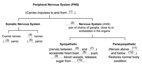
Solution E.3.
Fill in the following information in the diagram.
1. Central Nervous System
2. Autonomic
3. 12
4. Spinal
5. 31
6. Neck
7. Waist
8. dilates
9. constricts
10. liver
11. neck
12. sacral
Exercise 8.2
A.1.Which part of the eye is grafted in a needy patient from a donated eye?
(a) Conjunctiva
(b) Cornea
(c) Choroid
(d) Ciliary muscles
(a) Conjunctiva
(b) Cornea
(c) Choroid
(d) Ciliary muscles
(b) Cornea
A.2.Which part of our ear is shaped like a snail shell?
(a) Semi-circular canals
(b) Cochlea
(c) Stapes
(d) Eustachian tube
Solution A.2.
(b) Cochlea
A.3.The three parts of human ear contributing in hearing are:-
(a) Cochlea, ear ossicles and tympanum
(b) Semi-circular canals, utriculus and sacculus
(c) Eustachian tube, tympanum and utriculus
(d) Perilymph, ear ossicles and semi-circular canals
Solution A.3.
(c) Eustachian tube, tympanum and utriculus
A.4.The region in the eye where the rods and cones are located is the
(a) Retina
(b) Cornea
(c) Choroid
(d) Sclera
Solution A.4.
(a) Retina
B.1.Name the following:
(a) The photosensitive pigment present in the rods of the retina.
(b) The part which equalizes the air pressure in the middle and external ear.
(c) The ear ossicle attached to the tympanum.
(d) The tube which connects the cavity of the middle ear with the throat.
(e) The part of the eye responsible for its shape.
(f) The nerves which transmit impulse from ear to the brain.
(g) The photoreceptors found in the retina of the eye.
(h) The eye defect caused due to shortening of the eye ball from front to back.
Solution B.1.
(a) Rhodopsin
(b) Eustachian tube
(c) Hammer
(d) Dura mater
(e) Eustachian tube
(f) Cornea
(g) Auditory nerves
(h) Rods and cones
(i) Hypermetropia
B.2.Note the relationship between the first two words and suggest the suitable word/words for the fourth place.
(a) Cones: Iodopsin:: rods: ________.
(b) Sound: ear drum:: dynamic balance: __________.
Solution B.2.
(a) Cones: Iodopsin:: rods: rhodopsin
(b) Sound: ear drum:: dynamic balance: semi-circular canals
B.3.
Solution B.3.
C.1.Differentiate between members of each of the following pairs with reference to what is given in brackets.
(a) Myopia and hyperopia (cause of the defect)
(b) Rods and cones (sensitivity)
(c) Semi-circular canal and cochlea (Function)
(d) Rod and cone cells (pigment contained)
(e) Dynamic balance and static balance (definition)
Solution C.1.
(a) Myopia results when the eye ball is lengthened from front to back or the lens is too curved.
Hyperopia results from either too shortening of the eyeball from front to back or when the lens is too flat.
(b) Rods are sensitive to dim light but do not respond to colour.
cones are sensitive to bright light and are responsible for colour vision.
(c) cochlea is responsible for hearing; it can perceive the senses of hearing.
Semicircular canals are responsible for perceiving the senses to maintain the body balance.
(d) Rod cells contain rhodopsin whereas the cone cells contain iodopsin.
(e) Dynamic balance is when the body is in motion whereas static balance is positional balance with respect to gravity.
C.2.State whether the following statements are true (T) or false (F). If false, correct it by changing any one single word.
(a) Deafness is caused due to rupturing of the pinna.(T/F)
(b) Semicircular canals are concerned with static (positional) balance. (T/F)
C.2.State whether the following statements are true (T) or false (F). If false, correct it by changing any one single word.
(a) Deafness is caused due to rupturing of the pinna.(T/F)
(b) Semicircular canals are concerned with static (positional) balance. (T/F)
Solution C.2.
(a) False
Correct statement: Deafness is caused due to rupturing of the eardrum.
(b) False
Correct statement: Semicircular canals are concerned with dynamic balance.
C.3.Mention, where in living organisms are the following located and state their main functions:
(a) Fovea centralis
(b) Organ of corti
Solution C.3.
(a) Fovea centralis is located at the back of the eye almost at the centre of the eyeball. It is the region of the brightest vision and also of the colour vision.
(b) Organ of corti is located in the inner ear. It contains sensory cells which process hearing.C.4.Mention if the following statements are true or false. Give reason.
(a) Sometimes medicines dropped into the eyes come into the nose and even throat.
(b) Ciliary muscles regulate the size of the pupil.
(c) Yellow spot of the retina is the region of the colour vision.
(d) The auditory nerve is purely for perceiving sound.
(e) Malleus, incus and stapes are collectively called ear ossicles.
(f) Short-sightedness and hyperopia are one and the same thing.
(g) Blind spot is called so because no image is formed on it.
Solution C.4.
(a) True
(b) False/ Ciliary muscles regulate the size of the lens.
(c) True
(d) False/The auditory nerve responsible for sound as well as for the body balance.
(e) True
(f) False/ flavour is a combination of taste and smell.
(g) False/ short-sightedness is myopia and hyperopia is long-sightedness.
(h) True
C.5.Given below are two sets (a) and (b) of five parts in each. Rewrite them in correct sequence.
(a) Cochlea, tympanum, auditory canal, ear ossicles, oval window
(b) Conjunctiva, retina, cornea, optic nerve, lens
Solution C.5.
(a) Auditory canal, tympanum, ear ossicles, oval window, cochlea
(b) Conjunctiva, cornea, lens, retina, optic nerve
C.6.Given below are certain structures. Write against them their special functional activity.
(a) Organ of Corti and __________
(b) Olfactory nerve and ___________
(c) Retina and __________
Solution C.6.
(a) Organ of Corti and hearing
(b) Olfactory nerve and smell
(c) Retina and vision
C.7.Answer the following:
(a) What is the function of the lacrimal gland and where is it situated in the body?
(b) In what two ways is the yellow spot different from the blind spot?
(c) Name an age-old eye defect. What causes it?
(d) What is meant by the power of accommodation of the eye?
(e) Mention the characteristics of the image that falls on the retina of the eye.
Solution C.7.
(a) Lacrimal gland is a tear gland located at the upper sideward portion of the eye orbit. Its secreation lubricates the surface of the eye, washes aways the dust particles and kills germs
(b) Yellow spot is the region of brightest vision and contains maximum sensory cells whereas a blind spot contains no sensory cells and this is the point of no vision.
(c) Presbyopia is an age-old eye defect. In this condition, the lens loses flexibility resulting in far-sightedness.
Cataract is also very common in old people, the cornea becomes opaque and the vision is cut down even to blindness.(d) The process of focusing the eye at different distances is called the power of accommodation.
(e) The image formed on the retina is inverted and real.
C.8.What is meant by optical illusion? Give one example.
Solution C.8.
An optical illusion is the life-like continuous movement on the screen. Television is an example of optical illusion, where the scanning beam of a picture frame of the TV camera moves so rapidly on the viewing screen of the TV set that our eyes cannot keep pace with it.
C.9.Where are the following located? Briefly mention the function of each:
(a) Oval window
(b) Cochlea
(c) Semicircular canals
(d) Utriculus
C.9.Where are the following located? Briefly mention the function of each:
(a) Oval window
(b) Cochlea
(c) Semicircular canals
(d) Utriculus
Solution C.9.
(a) Oval window is located in the middle ear. It helps in setting the fluid in the cochlear canals into vibration.
(b) Cochlea is located in the inner ear. It helps in transmitting impulses to the brain via the auditory nerve.
(c) Semicircular canals are located in the inner ear. These help in maintaining the dynamic equilibrium of the body.
(d) Utriculus is located in the inner ear. It joins the semi-circular canals to cochlea. It also helps in maintaining static balance of the body.
C.10.
Solution C.10.
C.11.Complete the following table by filling in the blank spaces.
Structure
|
Function
|
1. Yellow Spot
|
(i) …………………….
|
2. …………………….
|
(ii) Transfers impulse from inner ear to brain
|
3. …………………….
|
(iii) Helps to change the focal length of the eye lens
|
4. Oval window
|
(iv) …………………….
|
5. …………………….
|
Dynamic equilibrium
|
Solution C.11.
D.1.Describe the mechanism of focusing the image of a distant object in your eye when you raise your head after reading a book.
Solution D.1.
While reading a book, the lens is more convex or rounded due to contraction of ciliary muscles because the book is usually read from a short distance. When we raise our head and look at a distant object, the ciliary muscles relax to build the tension on the suspensory ligament so that they can stretch the lens. This change in the curvature of the lens makes us focus on distant object.
D.2.Sometimes you remember a vivid picture of a dream you saw. What is the role of your eyes in this experience?
The brain sees the vivid picture of the dream through the eyes. Our eyes have actually never seen the vivid picture. This is an example of optical illusion. The area of dream is controlled by the cerebrum of the central nervous system. So sometime we can remember the vivid picture seen in the dream.
D.3.By closing the eyes and gently pressing them with your palms, you may see some specs of brilliant light. How do you get this sensation while there is no light entering your eyes?
Solution D.3.
If we look at a bright object and then close our eyes, the sensation of light persists for a short period. This is known as persistence image or the after image. It lasts for one-tenth of a second. Therefore by closing the eyes and gently pressing them with your palms, you see some specs of brilliant light.
D.4.Explain the terms 'adaptation' and 'accommodation' with reference to the eye.
Solution D.4.
Adaptation is the ability to adjust vision in bright and dark areas. When we enter a dark room from bright light, the rhodopsin pigment broken down in bright light is regenerated. It dilates the pupil and allows more light to enter the eyes. This is called dark adaptation. On the other hand, if we enter bright area from a dark room, the rhodopsin pigment is bleached. This constricts the pupil and reduces the light entering the eyes. This is called ‘light adaptation‘
Accommodation is the process of focusing the eye at different distances. This is mainly brought about by a change in the curvature of the lens. When the ciliary muscles contract, the lens becomes thicker and we are able to focus a nearby object. On the other hand when the ciliary muscles relax, the lens remains stretched i.e. the normal condition and we are able to focus on distant object.
D.5.You do not enjoy watching a movie from a very short distance from the screen in a cinema hall. Why?
Our eyes are designed to focus at a great variety of distances. To focus constantly at a short distance can make the lens focusing muscles fatigued. Therefore, we do not enjoy watching a movie from a very short distance from the screen in cinema hall.
D.6.Enumerate the common defects of vision, their causes and the possible methods of correcting them.
Solution D.6.
D.7.Name the three ear ossicles. How do they contribute in the mechanism of hearing?
Solution D.7.
The three ear ossicles are: Malleus (hammer), Incus (anvil) and Stapes (stirr up).
The last ear ossicle, stapes, vibrates and transmits the vibration to the oval window.
The role of other two ear ossicles is to magnify the vibration of stapes as a result of their lever like action.
D.8.What is meant by power of accommodation of the eye? Name the muscles of the eye responsible for the same.
Solution D.8.
The process of focusing the eye at different distances is called the power of accommodation. The ciliary muscles are responsible for the power of accommodation.
E.1.With reference to the functioning of the eye, answer the questions that follow:
a. What is meant by power of accommodation of the eye?
b. What is the shape of the lens during (1) near vision (2) distant?
c. Name the two structures in the eye responsible for bringing about the change in the shape of the lens.
d. Name the cells of the retina and their respective pigments which get activated (1) in the dark and (2) in the light.
Solution E.1.
a. The ability of the eye to focus sharply on things which are near to the eye as well as far off is known as the power of accommodation.
b. Shape of the eye:
Near vision – flattened
Distant – rounded or more convex
c. Ciliary muscles and suspensory ligament
d. In the dark: Cells – rod cells, Pigment – rhodopsin
In the light: Cells – cone cells, Pigment – iodopsin
E.2.With reference to the human ear, answer the questions that follow:
a. Given the technical term for the structure found in the inner ear.
b. Name the three small bones present in the middle ear. What is the biological term for them collectively?
c. Name the part of the ear associated with (1) static balance (2) hearing (3) dynamic balance.
d. Name the nerve, which transmits messages from the ear to the brain.
Solution E.2.
a. The middle ear or membranous labyrinth has two structures inside it, the cochlea and the semi-circular canals.
b. Malleus, incus and stapes
c. Static balance – Utriculus and sacculus (inner ear)
Hearing – Internal ear
Dynamic balance – Semi-circular canals (inner ear)
d. Collectively they are termed as ossicles.
E.3.The figure below compares a part of our eye with a part of a photographic camera.
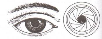
(a) Name the corresponding parts of the eye and the camera shown here that are comparable in function.
(b) Explain the mode of working and the functions of the parts of the eye mentioned above.
Solution E.3.
(a) Cornea is comparable to the lens cover of the camera.
The iris and pupil act like the aperture of a camera.
(b) The cornea is the eye’s main focusing element. It takes widely diverging rays of light and bends them through the pupil; the rays are further converged by the lens.
E.4.Given below is a diagram depicting a defect of the human eye? Study the same and answer the questions that follow:
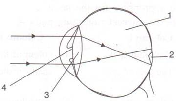
(a) Name the defect shown in the diagram.
(b) Give two possible reasons for this defect.
(c) Name the parts labeled 1 to 4.
(d) Name the type of lens used to correct this eye defect.
(e) Draw a labeled diagram to show how the above mentioned defect is rectified using the lens named above.
(a) Myopia
(b) The two possible reasons for myopia are either the eye ball is lengthened from front to back or the lens is too curved.
(c) 1 – vitreous humour, 2 – blind spot, 3-lens, 4-pupil
(d) Concave lens
E.5.The figure below is the sectional view of a part of the skull showing a sense organ:
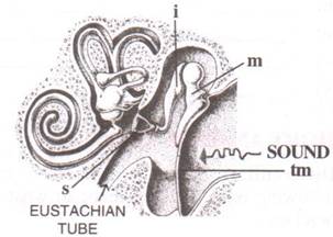
(iii) What do you call the part shown in the form of a spiral? What is its function?
(iv) Name the part labeled 'tm'. What is its function?
Solution E.5.
(i) Ear
(ii) m – malleus, i – incus and s – stapes respectively. These are collectively called as ear ossicles.
(iii) Cochlea. The vibrating movements in the hair of the sense cells of cochlea transmit the impulse for hearing to the brain via auditory nerve.
(iv) Tympanic membrane. It vibrates and then sets the ear ossicles into vibration in the process of hearing.
E.6.Given below is a diagram of a part of the human ear. Study the same and answer the questions that follow:
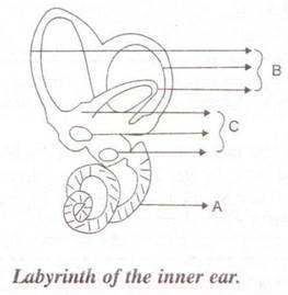
(i) Give the collective biological term for Malleus, Incus and Stapes.
(ii) Name the parts labeled A, B and C in the diagram.
(iii) State the functions of the parts labeled 'A' and 'B'.
(iv) Name the audio receptor region present in the part labeled 'A'.
(i) Ear ossicles
(ii) A – Cochlea, B – Semicircular canals, C – Ear ossicles
(iii) Cochlea helps in transmitting impulses to the brain via the auditory nerve. Semicircular canals help in maintaining dynamic equilibrium of the body.
(iv) Organ of Corti
E.7.Draw a labeled diagram of the inner ear. Name the part of the inner ear that is responsible for static balance in human beings.
Solution E.7.
E.8.Have a look at the posture of this woman who is reading a book and answer the questions which follow:
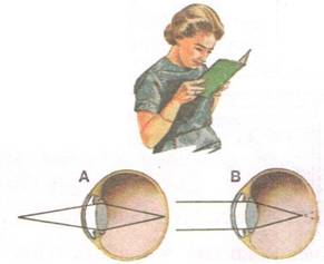
(a) What problem is she facing? Name the problem.
(b) What are the two conditions shown in sections A and B of the eye as applicable to her?
(c) What kind of looking glasses she needs?
(a) Myopia
(b) A-Normal eye, B-Myopia
(c) Looking glasses with the concave lens are required here.


